|
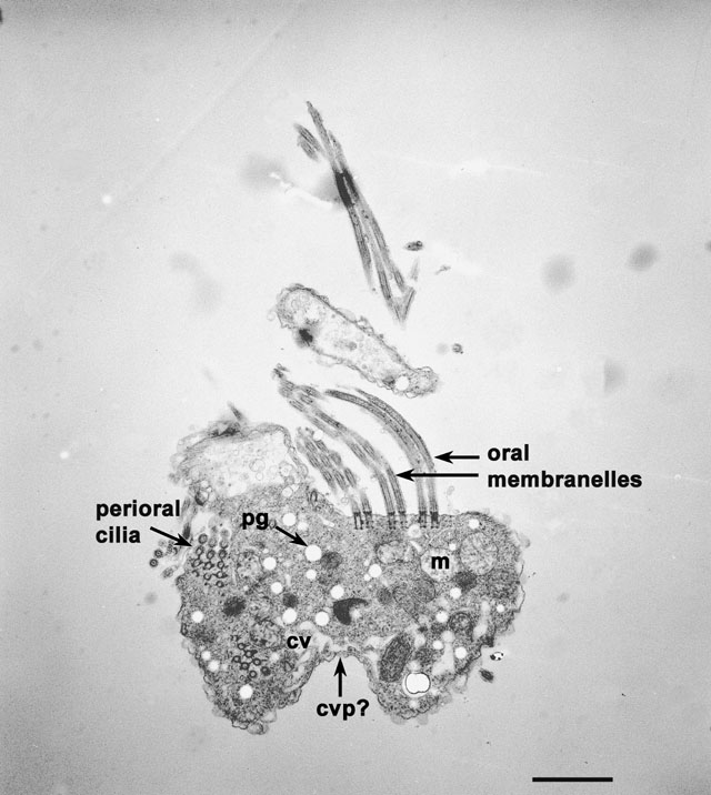
Halteria grandinella (obtained from Carolina Biological Supply
Co., NC, U.S.A.) is an oligotrich ciliate. This cell lives in fresh
water. It lacks long rows of somatic cilia called kineties but has a
few somatic ciliary bristles as well as a well developed oral and
adoral or perioral ciliature. The first set of 9 electron micrographs
is of a serially sectioned cell fixed in collidine-buffered fixative.
The sections are thicker than normal so that the EMs will appear less
sharp when enlarged. Images for enlargement are not provided for the
first 9 pictures. For a more complete study of this cell refer to
Grain, Protistologica 8:179-197, 1972. Figure 1a is of a section near
the cell’s left adoral surface where the contractile vacuole
(cv) is located and where some of the oral ciliature are
exposed in longitudinal section. A surface indentation may be near the
CV pore (cv p), (also see Fig. 9 below.) One membranelle of the
perioral ciliature is sectioned through its basal bodies. The cytosol
contains condensed as well as expanded mitochondria (m). The
uniformly round and electron transparent structures (pg) seen
here appear filled with dense material under different fixation and
staining conditions (see Fig. 2). These may be pigment granules
(pg). EM taken on 3/12/71 by R. Allen with Hitachi HU11A TEM.
Neg. 3,800X. Bar = 2µm.
|