 |
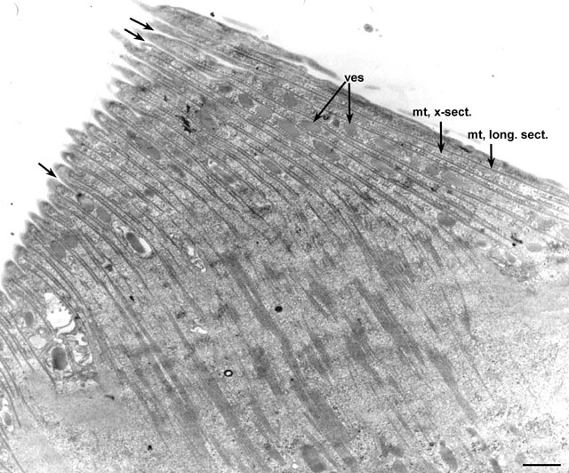
| Another view of the cytopharynx but in a dividing Didinium. This cytopharynx is in the proter (anterior) daughter cell. The lamellae, each consisting of a set of perpendicularly arranged microtubules (mt), cover this food-vacuole forming region. Vesicles (ves) lie near the cytopharyngeal membrane and fuse with the membrane (arrows). EM taken on 5/20/69 by R. Allen with Philips 300 TEM. Neg. 6,370X. Bar = 1µm. |
| Download High Resolution TIF Image (6.1 MB) |