|
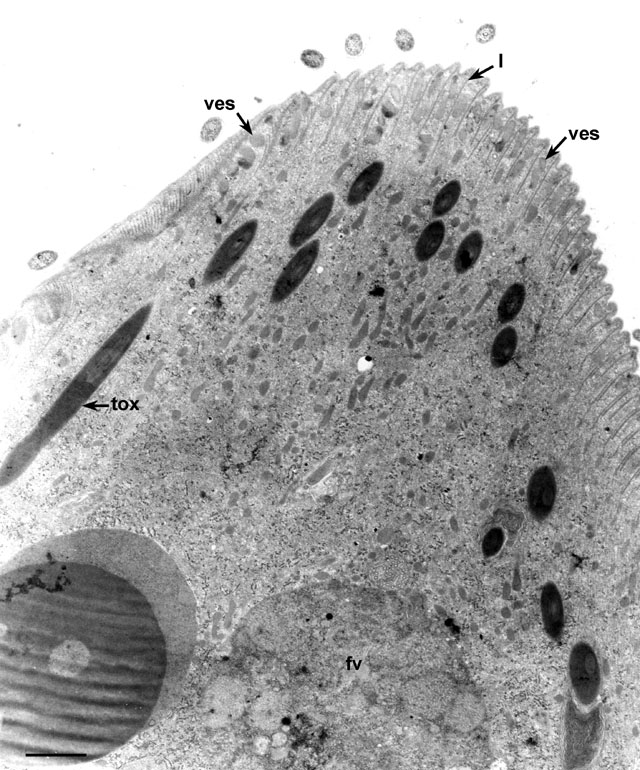
Didinium (non-dividing) has a massive cytopharynx at its
anterior end that takes the shape of a cone-shaped proboscis when
closed. This cytopharynx contains numerous lamellae (l)
composed of sets of microtubules (mt) that lie perpendicular to
each other next to the cytopharyngeal membrane. Vesicles (ves)
containing a uniform medium dense material (mucocysts?), line up
between the lamellae. Presumably these vesicles fuse with the single
cytopharyngeal membrane that covers the lamellae (arrows).
Extrusomes called toxicysts (tox) lie within in the
cytopharyngeal region. fv, food vacuole. EM taken on 9/19/68 by
R. Allen with Philips 300 TEM. Neg. 10,300X. Bar = 0.5µm.
|