|
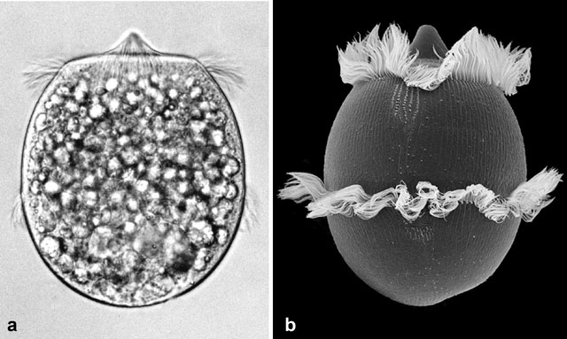
a) Phase contrast light micrograph of Didinium. This
barrel shaped ciliate is a predator feeding on Paramecium.
b) Scanning electron micrograph of Didinium showing the
two trochal bands of cilia surrounding the cell. The cilia show
metachronal waves produced by each cilium beating slightly out of
phase with the adjoining cilia. Patches of clavate (club-shaped) cilia
appear on this side of the cell. The cone-shaped cytopharynx is closed
at the cell’s anterior pole. Photographs courtesy of Dr. Klaus
Hausmann, Berlin, Gernmany.
|