|
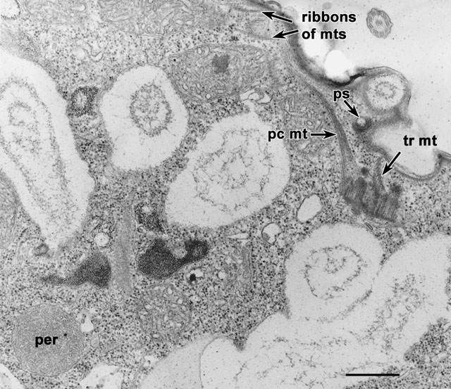
A tangential view through a dikinetid shows two (pc mt and
tr mt) microtubular ribbons extending toward the pellicle, one
from each basal body, and how they overlap with ribbons from other
basal bodies under the pellicle. The peroxisome (per) has one
membrane but is filled with numerous tiny circular profiles that may
represent tiny tubules. These tubules are much smaller than the
membranous cristae in the mitochondria and are more probably
proteinaceous in composition. EM taken on 6/3/69 by R. Allen with
Philips 300 TEM. Neg. 14,800X. Bar = 0.5µm.
|