|
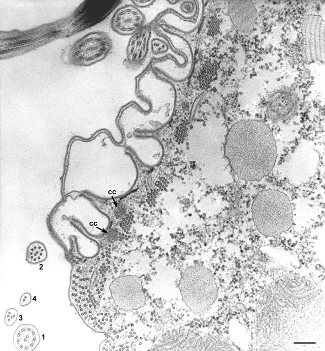
Detail of the junction between the ribbed wall and the right side of
the cytopharynx area similar to that at the top of Figure 24. The
ribbed wall is covered by alveoli and the ribs are produced by small
narrow ridges similar to the larger ridges covering the whole cell
body. Two arrays of microtubules are present in this zone, clumps of
hexagonally packed bundles lying under the ridges and rows of
microtubules that lie under the single cytopharyngeal membrane. There
is some suggestion that these rows of microtubules may associate
together to form the bundles. Tips of cilia in cross section give 4
views of how the tips of cilia end, the 9-doublet (1) first
become 9 singlets (2) and then these are lost one by one
(3) leaving only the two central singlets (4) at the tip
of a cilium. Sections of the cytostomal cord (cc) are also
present under the rib wall. This is the cord that wraps around the
cytopharynx as seen in 7b of figure 26 of Chapter 1. EM taken on
7/27/73 by R. Allen with Hitachi HU11A TEM. Neg. 23,000X. Bar = 0.2µm.
|