|
Paramecium caudatum is a peniculine oligohymenophoran ciliate
that has numerous rows of cilia called kineties that run along the
posterior to the anterior axis on the cell’s dorsal side but curve
around the oral region on the cell’s ventral side and end to form the
anterior and posterior suture lines that extend from the oral region
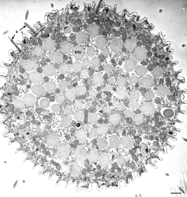
to the tips of the cell. Figure 1 is a cross-section of P.
caudatum near its anterior end showing a layer of trichocysts
(tric) that lie under the cell surface. The surface of the cell
is sculpted into ridges and the cilia (c) arise from the bases
of the concave invaginations. Each concavity represents a section
through one kinety so that in this micrograph approximately 58
kineties can be seen to surround the cell near its anterior end. At
this low magnification one can also see large numbers of medium-opaque
mitochondria near the cell surface and interspersed between the
trichocysts. Collidine-buffered fixation. EM taken on 1/18/74 by R.
Allen with Hitachi HU11A TEM. Neg. 3,750X. Bar = 1µm.
|