|
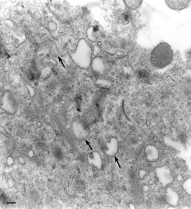
Figures 43-44 illustrate the changes in the CVC caused by cell
division and microinjecting mAbs specific for an antigen of the
V-ATPase holoenzyme into a P. multimicronucleatum cell. Each
condition causes the disappearance of the decorated tubules from the
radial arms. In this micrograph the cell was injected with 86 µg/ml of
serum containing the DS-1 mAb for the A4 antigen and lightly fixed 45
min later. It is observed that the bundles of decorated tubules had
started to round into vesicles (arrows), several tubules
forming one vesicle. By extrapolation, during bundle formation several
decorated tubules apparently arise from each vesicle which accounts
for the bundles of tubules emptying into a common duct. The common
duct opens into the smooth spongiome. We believe that the V-ATPases
have probably lost their functional integrity when the tubules
vesiculate as the tubules close to the vesicles in this micrograph no
longer have helical decorations. (See Ishida et al., J. Cell Sci.
108:693-702, 1993). EM taken on 3/30/93 by M. Ishida with Zeiss 10A
TEM. Neg. 19,800X. Bar = 0.2µm. Part published in J. Cell Sci.
108:693-702, 1993.
|