|
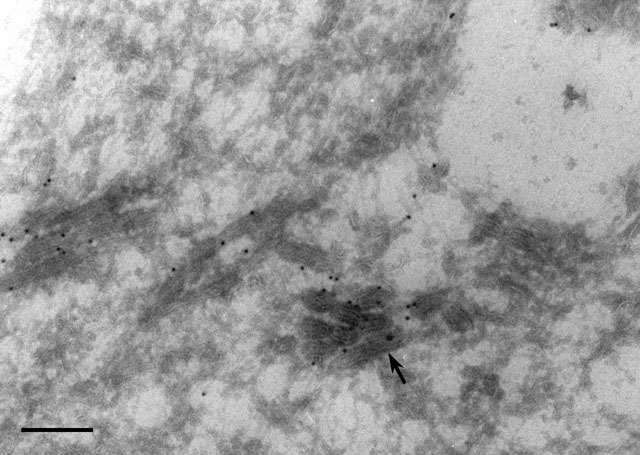
This micrograph shows the pegs on the surface of the decorated
tubules (arrow) that are negatively stained by the uranyl
acetate dissolved in the methylcellulose solution used to give support
to the frozen then thawed section. This section was labeled by a
polyclonal antibody to the V-ATPase of Dictyostelium which
cross-reacts with the V-ATPase in Paramecium (see Fok et al.,
J. Cell Sci. 108:3163-3170, 1995). EM taken on 2/28/92 by R. Allen
with Zeiss 10A TEM. Neg. 31,500X. Bar = 0.2µm. Small part published in
J. Cell Sci. 108:3163-3170, 1995.
|