|
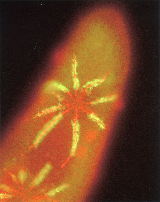
In addition to the mAb that specifically labels the A4 antigen of the
V-ATPase we also raised a mAb that labels an antigen, G4, of the
smooth spongiome. Since the mAb to A4 is a mouse IgG molecule and the
mAb to G4 is a mouse IgM molecule we were able to double label the
same cell using two different secondary antibodies, one tagged with
Texas red (G4) and the other tagged with fluorescein (A4). Double
labeling dramatically demonstrates that the smooth spongiome,
collecting canal, ampulla and contractile vacuole have the same G4
antigen (along with a pellicular membrane) while the decorated
spongiome alone bears the V-ATPase holoenzyme but not the G4 antigen.
This cell was recovering from a hyperosmotic stress so that the
concentration of decorated tubules was not yet as concentrated as in
control cells. Fluorescent micrograph taken by M. Iwamoto with Nikon
E400 Microscope and digital camera.
|