 |
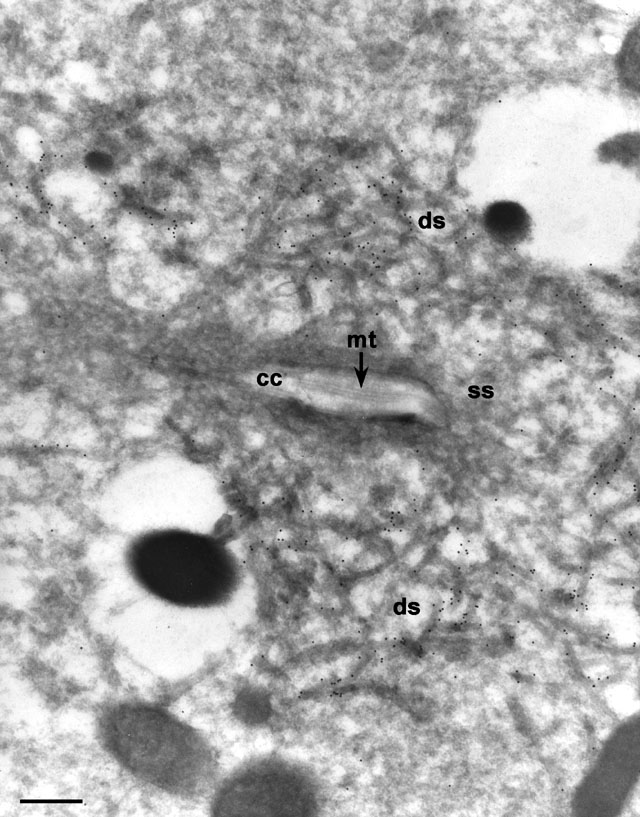
| When the antigen was visualized with immunogold using DS-1 on thin-frozen sections the antigen could be seen to be present on the periphery of the decorated tubules (ds). The collecting canal (cc) and smooth spongiome (ss) were not labeled with this mAb. mt, microtubules. EM taken on 3/22/86 by R. Allen with Zeiss 10A TEM. Neg. 12,000X. Bar = 0.5 µm. Part published in J. Cell Sci. 109:229-237, 1996. |
| Download High Resolution TIF Image (4.2 MB) |