|
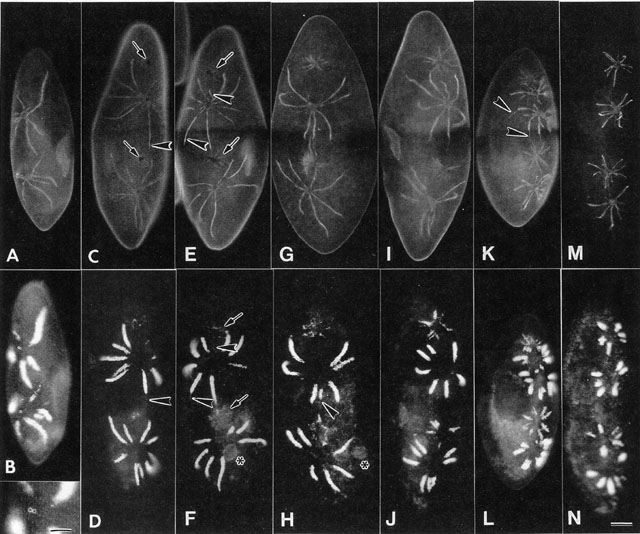
Double labeling experiments using mAbs to show the G4 antigen (SS-1
mAb) on the smooth spongiome (upper rows of pictures) and A4 antigen
on the decorated spongiome labeled by DS-1 (lower rows of pictures) of
the same cells show that the collecting canals also shorten during
division but sometimes at a slower rate (arrowheads). Thus it
is not just a loss of decorated tubules from around the distal ends of
the radial arms that occurs but all parts of the radial arms shorten
during cell division. Also during cell division there is a band around
the equator of the cell where the G4 antigen in the pellicle is
reduced in intensity and patches of reduced G4 antigen on the surface
of the cells (arrows) are the first indication of the sites
where the new CVCs will develop. A third mAb was used in B to
specifically label calmodulin. Calmodulin is found particularly around
the CV pore. Pictures taken by M. Aihara. Bar = 20µm. Published in J.
Eukaryot. Microbiol. 49:185-196, 2002.
|