 |
|
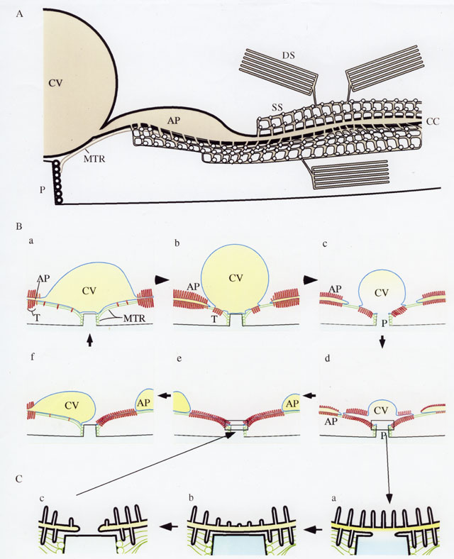
A. A small segment of the CVC of P. multimicronucleatum. The
edge of the contractile vacuole (CV) is next to an edge of the
CV pore (P). An ampulla (AP) is continuous with the CV
and the collecting canal is continuous with the ampulla. Smooth
spongiome (SS) in the form of cubic membrane of a gyroid
pattern opens into the collecting canals at intervals of 80nm between
the longitudinally segmented microtubular ribbons (MTR). Beyond
the ampulla bundles of 50nm-diameter tubules of the decorated
spongiome (DS) empty by common ducts into the smooth
spongiome. B. a) The CVC cycle consists of fluid flowing from the ampulla (AP) into the CV during diastole. Smooth spongiome tubules (T) of 40nm or cubic in form surround the ampullae and bind the CV to the microtubular (MTR) ribbons. b) Just before systole the CV rounds up and breaks away from the ampullae. c) The CV membrane fuses with the plasma membrane at the bottom of the CV pore and the CV membrane begins to collapse into tubules. Ampullae begin to fill. d) All of the CV tubulates against the microtubular ribbons and the pore can now close (see C). e) The pore closes. f) The filled ampullae can now fuse with the collapsed CV membrane, one ampulla at a time. Return to a) completes the cycle. C. The closure of the pore may require three steps: a) an iris diaphram like movement of the plasma membrane across the bottom of the pore sealing off the CV membrane, b) The release or fission of the CV from the pore membrane and c) fission of the CV membrane along each ribbon resulting in only one membrane, the plasma membrane, covering the bottom of the pore. This accounts for some electron micrographs showing only one membrane covering the pore during early diastole. This drawing was published as Figure 8A-C in Tominaga, Naitoh and Allen, J. Cell Sci. 112:3733-3745, 1999. |
| Download High Resolution TIF Image (20.1 MB) |