|
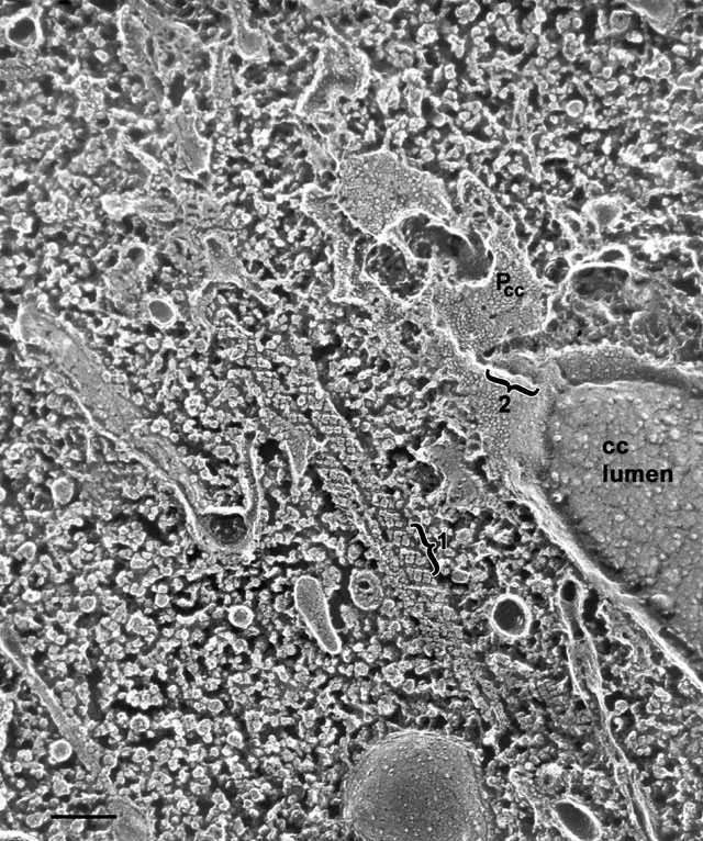
Quick-freeze deep-etch image of two decorated tubules with helically
wound subunits on their cytosolic surfaces. Each step in a helical
turn appears to be formed of three dyads (under bracket 1)
lying side by side across the row. Each dyad appears to be independent
(not fused) with the other two dyads at each step. Thus each helical
row seen in Fig. 27 apparently consists of a long series of three
dyads lying end to end rather than two rows of subunits as it was
assumed from Figure 20. This accounts for the three lines per helix
observed in some studies (McKanna, J. Ultrastruct. Res. 54:1-10, 1976
and in Fig. 20 above). Following the tubules downward in this picture
the fracture exposes the inside of one tubule and IMPs exposed here
may represent the luminal transmembrane extensions of their much
larger heads that appear on the cytosolic surface of the tubule.
Additionally, a filled collecting canal (cc) lies near the
decorated tubules with its P-fracture face (Pcc)
exposed that is studded with IMPs (where the luminal leaflet has been
removed). A row of indentations (bracket 2) leading to openings
to the smooth spongiome and holes (arrow) in a second row are
visible. IMPs appear to have different diameters on this P-face and
some are organized into rows. The content of the collecting canal
lumen has a uniquely etched pattern distinguishing it from other
vacuoles and the cytosol. EM taken on 6/22/88 by C. Schroeder with
Zeiss 10A TEM. Neg. 31,500X. Bar = 0.2µm. Published in J. Cell Sci.
108:3163-3170, 1995.
|