|
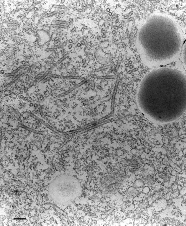
The decorated spongiome is composed of 50nm tubules that have a
pattern of helically wound subunits attached to their cytosolic
surfaces. These tubules can be up to one micrometer or more in length.
Each tubule ends blindly at one end but at its opposite end it empties
into a duct shared by several other tubules (as in Fig. 17). This
common duct empties into the smooth spongiome. EM taken 3/13/92 by R.
Allen with Zeiss 10A TEM. Neg. 19,800X. Bar = 0.2µm. Published in J.
Cell Sci. 108:3163-3170, 1995
|