|
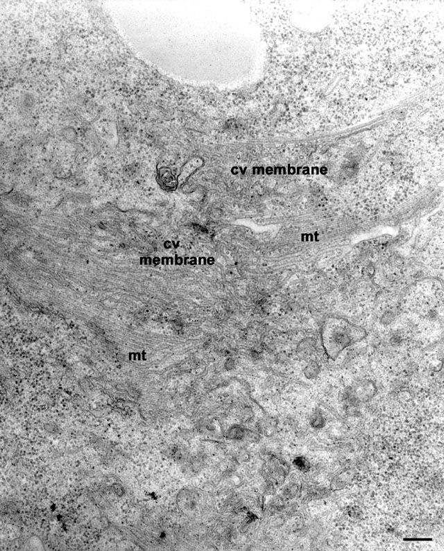
To determine what happens to the CV during systole we observed the CV
in a living cell and then fixed the cell as soon as the CV had opened
to the outside. It was apparent that the CV membrane does not flatten
into an empty sac but the CV membrane undergoes rapid tubule formation
and collapses as a continuous mat of tubules that maintains its
connection to the microtubular ribbons (mt). These tubules are
a uniform 40nm in diameter. EM taken on 6/9/96 by R. Allen with Zeiss
10A TEM. Neg. 19,800X. Bar = 0.2µm. Published in J. Exp. Biol.
200:1737-1744, 1997.
|