|
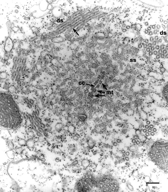
Normally the collecting canal is expanded when the CV is in systole.
In this micrograph the collecting canal is empty so that its two sides
are flattened against each other. Presumably the CV is in diastole
(filling phase). The ribbon of microtubules (mt) is split into
10 groups of two tubules each so the ribbon has 18 to 20 microtubules
at this level. The smooth spongiome (ss) is connected to the
collecting canal (seen better in unpublished serial sections) and
bundles of decorated spongiome (ds) form the peripheral layer
of the radial arm. The decorations consist of 2 closely set globular
units forming a helix around the tubule. In this micrograph the
globular units are outlined (arrow) with more electron dense
material so it appears there are 3 threads making up a helix. EM taken
on 12/9/70 by R. Allen with Hitachi HU11A TEM. Neg. 24,000X. Bar =
0.2µm.
|