|
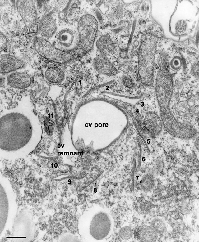
Figures 5 through 9 show a series of cross sections through one CVC
in late systole. Fig. 5 is a section through the CV pore close to the
bottom of the pore (#3 of a series of 43 sections). Eleven ribbons of
microtubules radiate from the pore in pin-wheel fashion and each
ribbon is bound to extensions of the CV membrane on its internally
slanted side. The section actually passes through the bottom of the
pore and exposes a small remnant of CV lumen that was not completely
expelled, to the lower left. The membrane next to the lowest ribbon
(ribbon #8) is visible as many cross-sectioned tubules. EM taken on
3/27/80 by R. Allen with Hitachi HU11A TEM. Neg. 12,250X. Bar = 0.5µm.
Published in J. Protozool. 35:63-71, 1988.
|