|
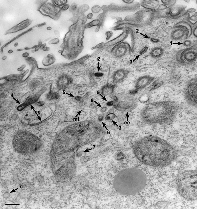
In a cell treated as in Figure 5, all stages of endocytosis and
movement to the early endosomes and out of the early endosomes
(ee) can be found. These steps are labeled 1 through 7 in
chronological order beginning at the parasomal sac and ending with
uncoated carrier vesicles ready to pass into the cell’s interior. This
micrograph is of the oral region. 1, coated pit of the
parasomal sac; 2, coated preendosomal vesicle; 3,
uncoated preendosomal vesicle; 4, fusion with early endosome;
5, budding coated pit from early endosome; 6, coated
carrier vesicles; 7, partially or totally uncoated carrier
vesicle. mt, microtubule. EM taken on 10/9/87 by R. Allen with
Zeiss 10A TEM. Neg. 19,800X. Bar = 0.25µm. Published in J. Cell Sci.
101:449-461, 1992.
|