|
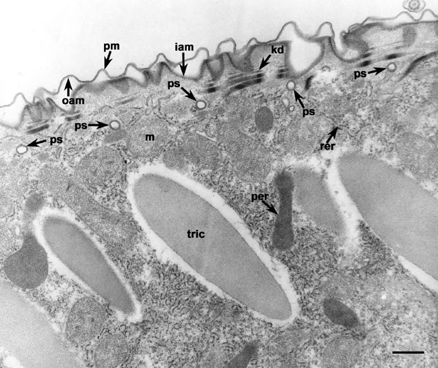
The pellicle of Paramecium consists of a plasma membrane
(pm) and alveolar sacs, with their inner (iam) and outer
(oam) alveolar membranes that limit the area in which
endocytosis can easily occur to only the parasomal sacs. Parasomal
sacs (ps) are cylindrical indentations of the plasma membrane
that pass through the alveoli, not by penetrating through the sac but
by passing through the septum. A short branch of the septum passes
around the parasomal sac. In some areas the parasomal sacs extend
almost perpendicularly from the basal bodies (not shown in this
micrograph). There is one parasomal sac per surface depression in
non-dividing cells. rer, rough ER; m, mitochondrion;
per, peroxisome; tric, trichocyst; kd,
kinetodesmal fiber stack. This cell was incubated in hydrogen peroxide
and diaminobenzidine to label the peroxisomes. EM taken on 2/27/81 by
R. Allen with Hitachi HU11A TEM. Neg. 10,250X. Bar = 0.5µm.
|