|
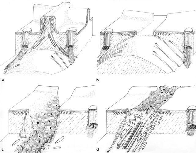
Drawing of the cytoproct illustrating four views as it changes shape
during vacuole defecation. a) A view of the short segment of
the closed and inactive cytoproct. Single microtubules extend into the
cytosol from the top of the ridge which is distinguished from other
ridges by piles of unique fibers. Bundles of microtubules also extend
inward from the adjoining basal bodies. b) The microtubules
contact the spent vacuole and move the vacuole toward the cell’s
surface. Ultimately the microtubules pull the vacuole and ridge apex
together where fusion between the vacuole and plasma membrane can
occur. c) Following fusion the spent vacuole membrane tubulates
and fission results in release of this membrane back into the cell.
Tubulation is promoted by actin filaments. d) The ridge will
reform when all spent vacuole membrane is retrieved. Published in J.
Cell Sci. 14:611-631, 1974.
|