|
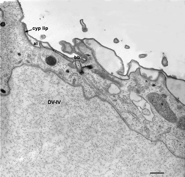
This cell had been treated with trifluoperizine which has the effect
of inhibiting the closure of the cytoproct. This drug must interfere
in some unknown way with the vesiculation of the membrane of the spent
vacuole. In this micrograph one lip of the open cytoproct (cyp)
is just visible in the upper left corner. However, another spent
vacuole (DV-IV) is approaching the open cytoproct and its
membrane is in contact with the microtubular bundle that arises from a
basal body (bb) next to the cytoproct. We conclude that motors
linking the microtubules to the vacuole membrane move the vacuole
toward the cell’s surface and towards its cytoproct. al,
alveolus. EM taken on 9/17/87 by R. Allen with Zeiss 10A TEM. Neg.
12,000X. Bar = 0.5µm.
|
