|
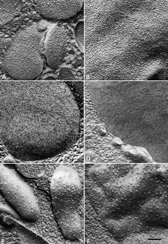
A comparison of conventional freeze fracture images of the E-fracture
faces of (A) discoidal vesicles with (B) the phagosome
membrane, (C) the acidosome membrane with (D) the
phagoacidosome membrane and (E) lysosome membrane with
(F) phagolysosome membranes. It is clear that these three
stages of digestive vacuoles (B, D and F) closely resemble the three
vesicle populations (A, C and E) that fuse with the digestive vacuole
immediately before that vacuole is transformed, respectively. This
plate was published as Figure 2 in Fok and Allen, In
Paramecium, H.-D. Gortz, ed., Springer Verlag, New York, pp. 301-324,
1988.
|