|
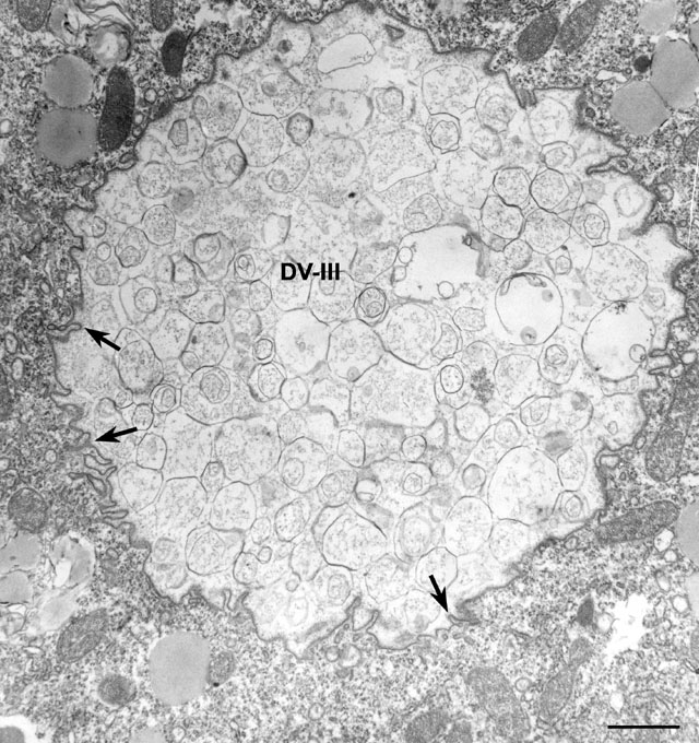
As with acidosome fusion with the DV-I, lysosome fusion with the
DV-II also seems to occur nearly all at once resulting in a
phagolysosome (DV-III) with a highly irregular surface. However,
unlike the transformation of DV-I into DV-II where the DV-I membrane
is removed by immunogold-labeled tubules we have very little
immunological evidence that the DV-II membrane is removed during the
transition from phagoacidosome to phagolysosome. Although there is
some morphological indication for tubular (arrows) and
vesicular formation around these early phagolysosomes (also see Fig.
50) that might indicate a similar transformation. The vacuole contents
seen here are thought to be the lipid micelles of the axenic medium.
EM taken on 2/27/80 by R. Allen with Hitachi HU11A TEM. Neg. 7,000X.
Bar = 1µm.
|
