|
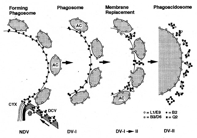
A summary of membrane replacement in the early steps of the digestive
vacuole cycle in P. multimicronucleatum. Six different mAbs
were raised to antigens found on subpopulations of membranes of the
digestive vacuole cycle. Discoidal vesicles (dcv) are labeled
luminally with mAb to B2 (filled circles) and on their
cytosolic surface with mAb for Q2 (filled squares). Discoidal
vesicles travel along the cytopharyngeal microtubular ribbons to the
cytopharynx (cyx) where they fuse with the nascent vacuole
membrane. Acidosomes (ac) are labeled luminally with mAbs to
both L1 and E9 antigens (open circles) and cytosolically with
mAbs to both B3 and D6 antigens. Acidosomes bind to the nascent
vacuole membrane and move with the phagosome to the cellís posterior
region. Here the acidosomes fuse with the phagosome to form the
phagoacidosome. Antigens L1/E9 and B3/D6 are transferred to the
phagoacidosome as the antigens B2 and Q2 are retrieved in tubules that
pinch off the maturing phagoacidosome. Thus the membrane of the
phagoacidosomes does not resemble the phagosome membrane in freeze
fracture morphology or in antigen content. Drawing published as Figure
12 in Allen et al., J. Cell Sci. 108:1263-1274, 1995.
|
