|
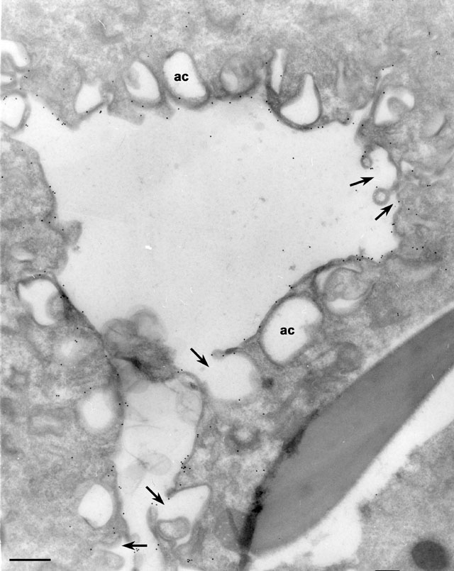
To label membranes inside the cell we used very lightly fixed cells
(0.25% glutaraldehyde) that were then rapidly frozen in liquid
nitrogen and sectioned later at -100oC. These frozen
sections were picked up on drops of methylcellulose and transferred to
a Formvar-supported grid. The sections were immunogold labeled (15nm
gold) to show the location of the specific antigen inside the cell as
well as on the cell surface. The same mAb used in Fig. 8a can be seen
here on the DV-I membrane that is detached from the cytopharynx. Some
acidosomes have already fused with this phagosome (arrows). The
unfused acidosomes (ac are not labeled with this mAb. EM taken
on 4/3/87 by R. Allen with Zeiss 10A TEM. Neg. 12,000X. Bar = 0.5µm.
Part published in J. Cell Sci. 108:1263-1274, 1995.
|