|
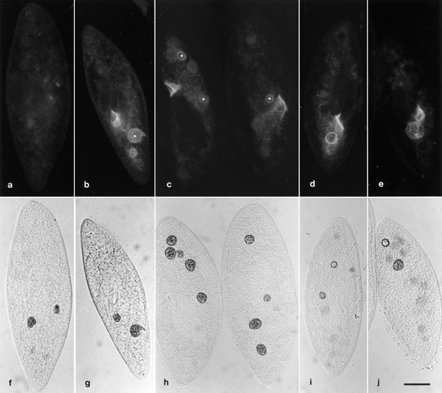
Monoclonal antibody for antigen B2 (called mAb PC-1 in Fok et al.,
Eur. J. Cell Biol. 40:1-8, 1986) is located on the cytopharynx and
along the cytopharyngeal ribbons, as well as on 1 or 2 recently formed
digestive vacuoles (DV-I). This was determined by feeding the cells
latex beads (PLS) in pulse-chase studies and comparing the cells'
images in fluorescent and in light microscopy. Fluorescent images a to
e are of the same cells as f to j, respectively. DVs containing PLS
are 1 min. old or less in a/f and b/g; 2-5 min. old in c/h; 6-9 min.
old in d/i; and 22-25 min. old in e/j. DVs (white dots) that
fluoresce are found only in cells up to 5 min. in age. Control cell
a/f was fed PLS but was not treated with mAb for B2. Pictures taken by
M. Aihara. Bar = 20µm.
|