|
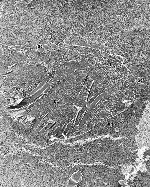
The nascent phagosome membrane has a dense coat of acidosomes
(ac) docked at its cytosolic surface well before the nascent
vacuole pinches off the cytopharynx. This micrograph is a quick-freeze
deep-etch image of a transverse section through the posterior portion
of the buccal cavity with a nascent vacuole bulging from the
cytopharynx (cp). Living cells were spun down in a centrifuge
and a slurry of cells was mounted on a copper support before slam
freezing the living cells against a highly polished copper surface.
Some paramecia had been disrupted and this cell was feeding on the
particulate organelles of the disrupted cells. Thus the nascent
vacuole contains these particles amassed against its luminal
(lu) surface. The cilia (c) in the buccal cavity
(bc) obviously efficiently sweep the particles against the
dorsal posterior surface. EM taken on 5/26/92 by R. Allen with Zeiss
10A TEM. Neg. 4,000X. Bar = 1µm. Published in J. Cell Sci.
106:411-422, 1993.
|
