 |
|
Development of the digestive vacuole or phagosome was discussed and
illustrated in the previous section as this is the function of the
cytopharynx after the food organisms or particles have been filtered
through the buccal cavity to its posterior dorsal surface (see Ishida
et al., J. Eukaryot. Microbiol. 48:640-646, 2001). This section will
show the changes that occur in the digestive vacuole as it passes
through the cell. The next section on the cytoproct will describe the
fate of the spent vacuole, both its content and membrane. For
convenience we will refer to the digestive vacuole stages as DV-I, II,
III and IV. DV-I is the phagosome, DV-II is the phagoacidosome, DV-III
is the phagolysosome and DV-IV is the spent phagosome or spent
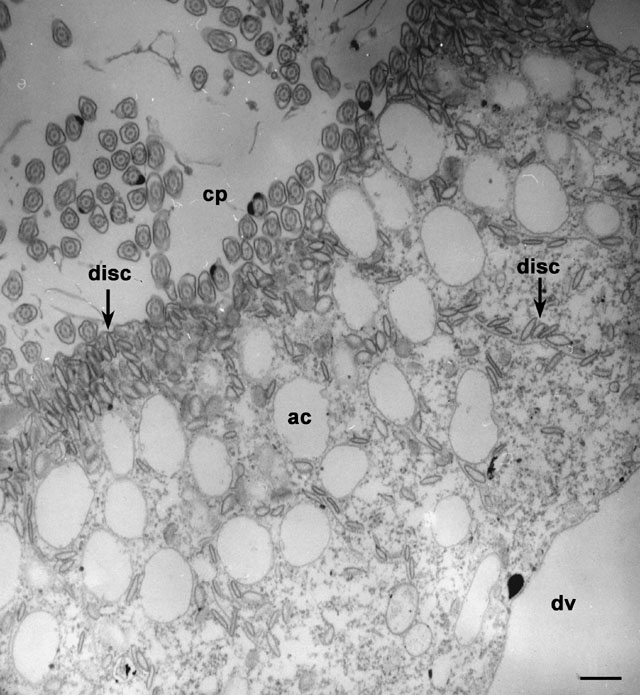
digestive vacuole. Two of the three types of vesicles that are transported to the cytopharynx (cp) and that accumulate in large numbers at the cytopharynx are shown in this electron micrograph. The discoidal vesicles (disc) move up to the edge of the cytopharynx membrane where the cytopharynx membrane is continuous with the plasma membrane of the buccal cavity. Here the discoidal vesicles fuse with the cytopharynx membrane. Just behind this edge is found a layer of larger vesicles, the acidosomes (ac), which dock with the cytopharynx membrane as soon as they make contact with this membrane. Unlike the discoidal vesicles these acidosomes do not fuse with the cytopharynx or nascent vacuole until the phagosome has pinched off and moved toward the posterior pole of the cell. EM taken on 4/8/83 by R. Allen with Hitachi HU11A TEM. Neg. 8,000X. Bar = 0.5µm. |
| Download High Resolution TIF Image (3.5 MB) |