 |
|
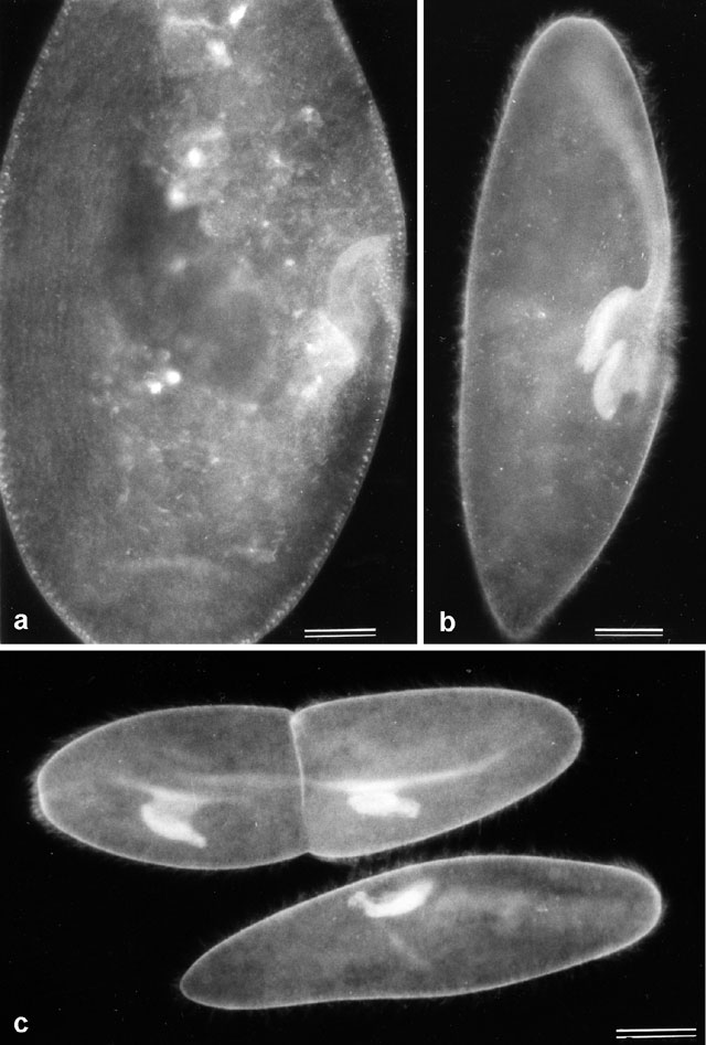
A. ConA-FITC labeled P. multimicronucleatum shows the true
shape of the buccal cavity, which is not quite as long as the clay
model in Figure 2b and its anterior curves less abruptly. The
cytopharynx (cp) is more fluorescent and curves more quickly
over the side of the buccal cavity than the clay model suggests. The
orientation of this image is similar to part lc of Figure 2b although
the depth of focus superimposes both right and left sides on top of
each other in this image so one cannot distinguish right from left
sides in this side view. Bar = 20µm. B. The new buccal cavity forms in association with the mature buccal cavity as it arises from a region of unorganized basal bodies called the anarchic field (see 1d in Figure 2b) that lies between the endoral membranelle and the vestibulum. Bar = 20µm. C. Later in division the two oral regions move apart as the cell elongates in its mid section and the division furrow forms. Pictures a to c taken by M. Aihara. Bar = 20µm. Published in J. Protozool. 35:400-407, 1988.
|
| Download High Resolution TIF Image (2.2 MB) |