 |
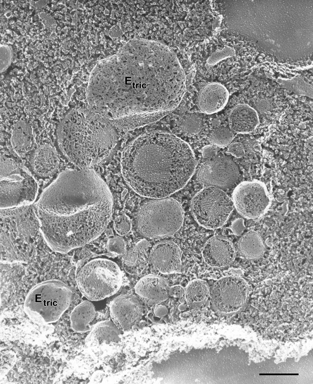
| Quick-freeze deep-etch image of a cytosolic region where trichocysts are developing. Vesicles, even small ones, contain contents that are compacting into paracrystalline form. These vesicles seem to have an affinity for each other as many are in contact. E-faces of the trichocyst membrane (Etric) are pitted to varying extents. The fracture plane reveals different planes of packing in the core. EM taken on 6/7/88 by C. Schroeder with Zeiss 10A TEM. Neg. 15,900X. Bar = 0.5µm. |
| Download High Resolution TIF Image (5.2 MB) |