 |
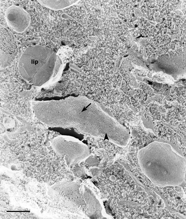
| Another view of the ER on which IMPs are organized into lines (arrowhead), arcs and circles (arrow) indicating organization of proteins within the ER membrane and possibly peripheral proteins attached to its cytosolic surface. lip, lipid. EM taken on 3/4/97 by R. Allen with Zeiss 10A TEM. Neg. 15,900X. Bar = 0.5µm. |
| Download High Resolution TIF Image (7.2 MB) |