|
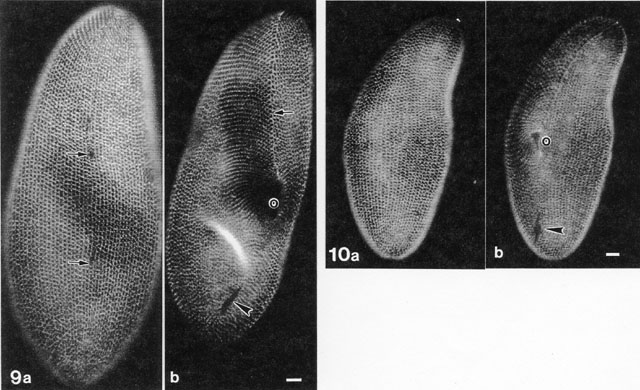
We raised a monoclonal antibody to the striated bands of P.
multimicronucleatum which is shown in the fluorescent micrographs
in Figures 9 and 10. The pattern of this cytoskeletal meshwork is
different from the infraciliary lattice shown in Fig. 26. The sites of
basal bodies and cilia are unlabeled and longitudinal rows of these
sites correspond to the kineties. The striated bands form a layer
under the longitudinal and lateral ridges of the pellicle. Contractile
vacuole pores appear in Fig. 9a (arrows) as unlabeled, areas
and the anterior suture along the ventral surface is apparent in Fig.
9b (arrow). Fig. 10a-b are of P. tetraurelia which
reacts with the same mAb demonstrating that striated bands are also
present in this species. arrowhead, cytoproct; o, oral
region. Micrographs taken by M. Aihara. Published in J. Euk.
Microbiol. 45:202-209, 1998.
|