|
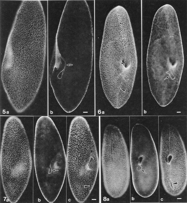
Fluorescent micrograph of centrin antibody labeling of the
infraciliary lattice of different species of Paramecium. This
plate was published as figures 5 to 8 in Allen, Aihara and Fok, J.
Eukaryot. Microbiol. 45:202-209, 1998. Figures 5a and 6a show a side
view and ventral view of P. multimicronucleatum. Figure 5b and
6b show the extension of the infraciliary lattice that encircles the
cytopharynx as the “cytostomal cord”. This cord is an extension of the
anterior suture cytoskeleton. Figures 7a-c are of P. caudatum
and Figures 8a-c are of P. tetraurelia. All pictures are taken
at the same magnification. arrowheads, cytoproct;
arrows, cytostomal cords. Anti-centrin antibody was a gift from
Dr. J. Salisbury. Micrographs taken by M. Aihara. Published in J. Euk.
Microbiol.45:202-209, 1998.
|