|
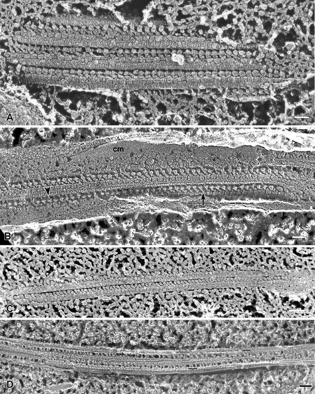
A. The outer dynein arms along the A subfiber appear as
globular units attached to the microtubule by stem-like connections.
The ciliary membrane was removed during the preparation and the
axoneme was probably in rigor so that all dyneins are bound to the
adjacent doublet. EM taken on 5/6/88 by C. Schroeder with Zeiss 10A
TEM. Neg. 40,500X. Bar = 50nm. B. QF-DE of a portion of an
isolated cilium exposing two rows of outer dynein arms arising from
subfiber A of each doublet. The heavy chains of the dynein complex are
linked to the microtubule by stems (arrow) that can be seen in
this micrograph. Thin protrusions (arrowhead) link the dynein
heavy chains to the adjacent doublet. Much of this cilium is still
covered by the ciliary membrane (cm). EM Taken on 6/7/88 by C.
Schroeder with Zeiss 10A TEM. Neg. 19,800X. Bar = 0.1µm. C.
QF-DE of another segment of the edge of the axoneme showing the outer
axonemal dynein arms that extend from the A-subfiber of a doublet to
bind to the B subfiber of the adjacent doublet (for cross-sectional
details of a cilium, see Chaper 11, figure 21). EM taken on 4/21/88 by
C. Schroeder with Zeiss 10A TEM. Neg. 31,500X. Bar = 0.1µm. D.
QF-DE of an isolated and longitudinally fractured cilium exposing the
middle of the cilium. The spokes extending from the doublets toward
the two singlet microtubules (see Chapter 11, figure 21) are viewed.
These spokes are spaced unevenly so they appear to be arranged into
triplets (brackets). The fracture plane, while in general is
longitudinal, exposes the singlets and outer doublets at different
levels so various periodicities appear along these microtubules. The
cilium may also be slightly twisted along its length. EM taken on
2/15/88 by C. Schroeder with Zeiss 10A TEM. Neg. 31,500X. Bar = 0.1µm.
|