|
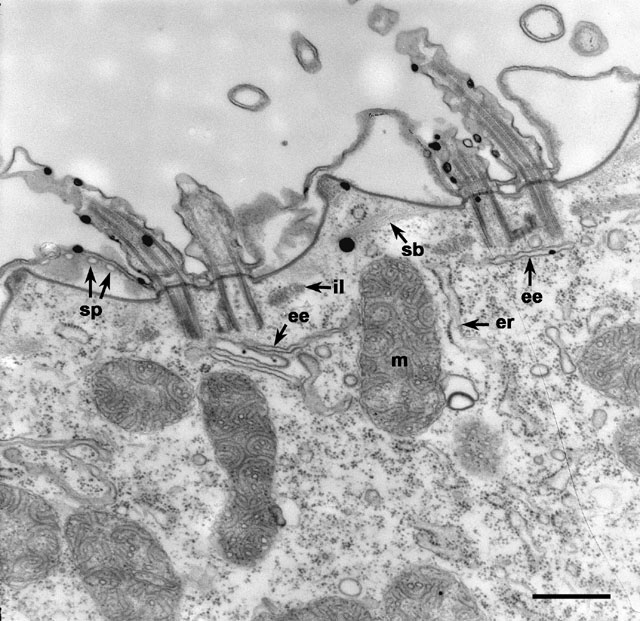
Two basal bodies, with attached cilia, arise from the bottoms of each
of two surface indentations of the cell surface. A partial septum is
observed to the left of the figure where the outer and inner alveolar
membranes join. This septum is perforated with pores (sp) next
to the outer alveolar membrane. Thus the lumens of adjacent alveoli
are continuous because of this system of pores. This cell has calcium
deposits (electron-opaque blobs) in its cilia and cortex as this cell
was exposed to calcium in its growth medium. il, infraciliary
lattice; sb, striated bands; m, mitochondrion;
er, endoplasmic reticulum; ee, early endosomes. EM taken
on 1/10/73 by R. Allen with Hitachi HU11A TEM. Neg. 16,900X. Bar =
0.5µm. Published in Poste and Nicolson (eds.), Membrane Fusion, p.125,
Elsevier/North Holland Biomed. Press, 1978.
|