 |
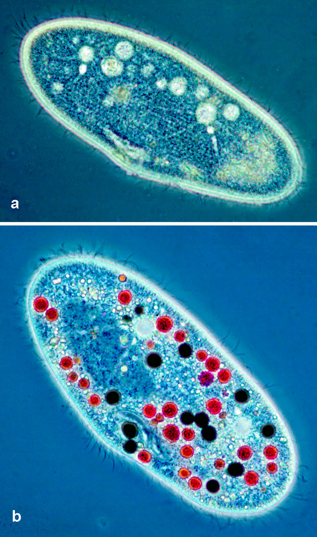
| a) Phase contrast light micrograph of Paramecium highlighting the two contractile vacuoles including the radial arms each composed of ampulla and collecting canal. b) Phase contrast micrograph showing food vacuoles in Paramecium stained either with carmine particles (red) or India ink (black). The cell is covered with cilia. Photographs courtesy of Dr. Klaus Hausmann, Berlin, Germany. For more pictures see http://microscope.mbl.edu. |
| Download High Resolution TIF Image (2.1 MB) |